In this interview, Claudia Florindo talks about what matters when comparing benchtop fluorescence microscopes.
What prompted Oxford Instruments to produce this buyer’s guide, and what gaps or recurring questions were you hoping it would address?
Over the last few years, we’ve seen a dramatic expansion in the capabilities available in benchtop fluorescence microscopy. What was once a relatively straightforward purchasing decision now involves choices between widefield, confocal, super-resolution, live-cell imaging, automation, image analysis, and a growing range of software and workflow options.
Many researchers come to us asking similar questions: Do I really need confocal? How important is detector sensitivity? What software features should I expect? How can I balance performance, usability, and budget? We created this guide to provide an objective framework for answering those questions.
Our goal was not to promote a particular technology, but to help users make informed decisions based on their applications, samples, workflows, and future requirements. We wanted buyers to understand that selecting a microscope is about much more than resolution specifications alone.
Image Credits: Oxford Instruments
Who is the guide primarily aimed at, and how did you account for readers ranging from experienced microscopists to those new to fluorescence imaging?
The guide was intentionally written for a broad audience. Benchtop microscopy is increasingly being adopted by experienced imaging specialists and researchers in biotechnology, pharmaceutical development, teaching laboratories, clinical research, and emerging start-ups.
For newcomers, we explain the fundamentals of imaging modes, optics, detectors, and workflow considerations without assuming extensive microscopy expertise. For more experienced microscopists, we discuss technical topics such as objective correction classes, detector quantum efficiency, confocal implementations, quality control frameworks, and upgradability.
The intention was to create a practical guide that could be useful whether someone is purchasing their first fluorescence microscope or replacing an existing imaging platform.
Benchtop fluorescence microscopes now cover a very broad range of capabilities. Why can choosing the right system be more complicated than it first appears?
The term “benchtop microscope” can be misleading because it describes the physical format rather than the instrument’s actual capabilities.
Benchtop systems vary in price and size, but even when they have a similar footprint, two systems may deliver dramatically different performance in imaging depth, sensitivity, acquisition speed, optical sectioning, automation, and software functionality. Some systems are primarily designed for routine imaging, while others support advanced 3D imaging, live-cell experiments, automation workflows, or even super-resolution techniques.
As a result, buyers who focus only on form factor, price, or benchtop risk overspecifying or underspecifying their needs. The real challenge is identifying which capabilities will add genuine value to the intended application and which features may never be used.
The guide covers imaging modes including transmitted light, widefield, confocal and super-resolution microscopy. What should buyers consider when deciding which of these capabilities they genuinely need?
The best starting point is always the biological question being asked.
For routine fluorescence imaging of relatively thin samples, widefield imaging often provides excellent results with high speed and low complexity. If users need optical sectioning, improved contrast in thick samples, or high-quality 3D reconstruction, confocal microscopy becomes increasingly important.
Super-resolution should be considered only when the scientific question genuinely requires information below the conventional diffraction limit. While super-resolution can reveal remarkable structural detail, it often comes with additional complexity, longer acquisition times, and increased costs.
A useful question to ask is: What decision or discovery will this additional resolution enable? If there is no clear answer, standard fluorescence imaging may be the more practical choice.
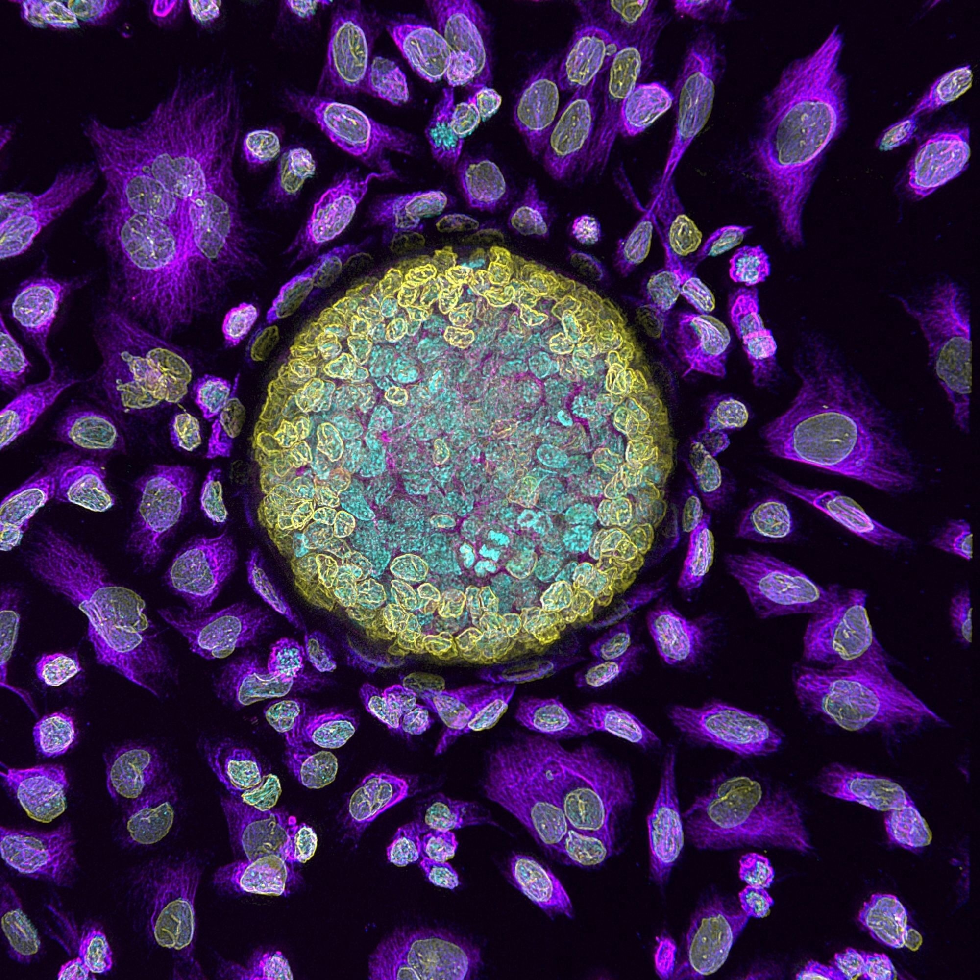 Image Credits: Dr Sebastian Amos and Dr Yu-Suk Choi, University of Western Australia
Beyond headline specifications such as resolution and magnification, which components or performance factors are most often overlooked during the purchasing process?
Software is probably the most underestimated component of a microscope purchase.
Researchers often focus on optics and resolution, but day-to-day productivity is heavily influenced by how easy it is to acquire, process, analyze, and share data. Features such as automated stitching, deconvolution, autofocus, multiposition imaging, and integrated analysis can significantly affect workflow efficiency.
Other commonly overlooked factors include detector sensitivity, field of view, stage accuracy, environmental stability, vibration control, service support, and quality control procedures. These may not appear in marketing headlines, but they have a major impact on the reliability and reproducibility of results over time.
How important is it for buyers to begin with their samples, applications and workflow requirements, rather than focusing immediately on a particular instrument or technology?
I would argue it is absolutely essential.
The starting point should always be the sample and the scientific question being asked. Thick organoids, live-cell cultures, tissue sections, small model organisms, and high-throughput screening applications all place very different demands on a microscope, and no single technology is the optimal solution for every application.
When buyers focus on a specific technology first, whether that’s confocal, super-resolution, or any other imaging modality, there is a risk of becoming distracted by impressive specifications that may not actually address their experimental needs. By starting with the sample, the biological question, and the practical workflow requirements, the technology choice tends to emerge much more naturally.
In our experience, the most successful microscope investments are made when researchers clearly define what they want to image, the level of detail they need to achieve, how they intend to acquire and analyze the data, and how frequently the system will be used. These factors are often more important than any individual specification on a datasheet.
How can researchers without extensive microscopy expertise make informed purchasing decisions?
It’s important to recognize that not every researcher is a microscopy expert, and they shouldn’t be expected to be. That’s where experienced sales engineers and application specialists play an important role. A good supplier should take the time to understand the user’s samples, applications, budget, and future ambitions, and then recommend the technology that will best support their research, rather than simply promoting the most advanced or expensive system. Ultimately, microscopy is about enabling scientific discovery, and the technology should serve the application, not the other way around.
The guide discusses practical considerations such as stage design, environmental control, software, automation, service, and quality control. How can these factors affect day-to-day usability and the long-term value of a microscope?
These factors often determine whether a microscope becomes a routinely used research tool or an underutilized asset.
For example, accurate motorized stages enable reproducible multi-position and large-area imaging. Environmental control becomes critical for live-cell and long-term experiments. Automation reduces user variability and improves consistency across laboratories. Software affects how quickly data can be acquired and analyzed.
Similarly, a strong service program and documented quality-control procedures help ensure that performance remains consistent over the instrument’s lifetime. In regulated or collaborative environments, the ability to verify system performance can be just as important as the imaging performance itself.
Image Credits: Oxford Instruments
Cost is clearly an important consideration. What advice would you give laboratories trying to balance current imaging needs with budget limitations and possible future requirements?
My advice would be to focus on value rather than simply acquisition cost.
A lower-cost system may appear attractive initially, but if it cannot support future applications or requires replacement after a few years, the overall cost of ownership can be much higher.
At the same time, laboratories should avoid paying for capabilities they are unlikely to use. The ideal solution is often a platform that addresses current requirements while allowing expansion as research evolves.
Carefully evaluating upgrade pathways, software scalability, service options, and application flexibility can help laboratories maximize the return on their investment.
Upgradability and multimodality feature prominently in the guide. Why are these becoming increasingly important when laboratories invest in benchtop imaging systems?
Research evolves quickly. The questions being asked today are often different from those that will be asked three or five years from now.
Many laboratories begin with straightforward fluorescence imaging but later require confocal imaging, advanced automation, live-cell workflows, or higher-resolution capabilities. A modular, upgradeable platform allows those new requirements to be addressed without replacing the entire system.
Multimodality is equally important because a single laboratory often works with multiple sample types. Having access to complementary imaging modes within one platform provides flexibility while simplifying training, maintenance, and workflow standardization.
After reading the guide, what are the three most important questions you would encourage a prospective buyer to ask a microscope supplier before making a final decision?
First: Can you demonstrate that the system performs well on my actual samples?
Nothing is more valuable than seeing real-world results generated using the specimens and workflows that matter to your research.
Second: What can this system become in the future?
Understanding upgrade pathways, software scalability, and long-term support helps protect the investment and avoids unnecessary future replacement costs.
Third: How will you help me maintain performance over the lifetime of the instrument?
This includes service support, training, application assistance, and quality control programs. A microscope purchase is not simply a hardware transaction; it is a long-term partnership that should support scientific success for many years.
As a final conclusion, if there is one message I’d like readers to take away from the guide, it’s that the best microscope is not necessarily the one with the most features. It’s the one that best matches your samples, experimental questions, workflow requirements, and future ambitions. Taking a structured approach to evaluation helps ensure that laboratories invest in a system that delivers value from day one and continues to support their science as it evolves.
About Claudia Florindo
Claudia Florindo is a Product Manager for Microscopy Systems, working closely with researchers and imaging specialists to advance microscopy workflows and data-driven scientific discovery. With more than seven years of experience in the scientific imaging industry and a strong background in life sciences research, she combines expertise in microscopy systems, imaging software, scientific cameras, and quantitative image analysis. Claudia collaborates with multidisciplinary teams across academia and industry to translate customer needs into innovative product solutions and support the adoption of advanced imaging technologies. Her experience spans product strategy, new product introduction, market analysis, and customer-driven product development, helping researchers maximize the value of their microscopy data through efficient, reproducible, and user-focused workflows. Through her work, Claudia plays a key role in driving product innovation and enabling the scientific community to accelerate discovery through advanced imaging solutions.
About Oxford Instruments – Life Sciences
Oxford Instruments is a leading provider of high-technology tools and systems for research and industry, dedicated to accelerating breakthroughs that create a brighter future for our world. With a global presence, we are committed to innovation and excellence, offering cutting-edge solutions that enable researchers and industry professionals to achieve breakthroughs in their fields. Our advanced technologies deliver numerous benefits through unparalleled precision and reliability, allowing users to obtain accurate and reproducible results. By utilizing Oxford Instruments’ innovative solutions, research is accelerated, productivity is enhanced, and innovation is achieved in various fields, including materials analysis, life sciences, semiconductors, physics, chemistry, and food sciences. We take pride in being a trusted partner for those aiming to push the boundaries of scientific and industrial advancements, providing the tools and support necessary to realize their visions.
Atomic force microscopy: These advanced instruments are utilized for high-resolution imaging and precise measurement of surface properties at the nanoscale level. They offer detailed topographical information and enable accurate measurements of features such as height, roughness, and mechanical properties.
Light microscopy: Our solutions encompass a comprehensive range of advanced imaging systems that utilize visible light for examining samples at the microscale. Equipped with high-quality lenses, cameras, and illumination systems, these optical microscopes deliver detailed images with exceptional clarity and resolution. They find applications in fields such as biology, materials science, and forensics.
Plasma etch & deposition: innovative etch & deposition techniques, atomic layer etch and atomic layer deposition, and ionbeam etch & deposition solutions used to produce high yielding, efficient and reliable advanced semiconductor devices. Our service and product range includes research and development systems right through to fully clusterable, robust and high up-time production systems, designed for high-volume manufacturing in key markets like quantum, power devices, augmented reality and datacom.
Electron microscopy analysis: Our high-performance tools are tailored for materials characterization, particle analysis, and sample manipulation at the nanometer scale. Techniques such as Backscatter Electron and X-ray (BEX), Energy Dispersive Spectroscopy (EDS), and Electron Backscatter Diffraction (EBSD) enable imaging, chemical analysis, and crystallographic characterization of materials at atomic and nanoscale levels.
Optical imaging and spectroscopy: Our optical imaging solutions comprise a range of advanced technologies and systems for capturing and analyzing images using light. From state-of-the-art optical microscopes to spectroscopy systems and imaging software, these solutions provide high-quality images and data for applications such as materials characterization, biological research, and semiconductor analysis.
Nanoindentation: Our high-resolution, MEMS-based nanoindenters are designed to measure the mechanical properties of materials, including hardness, elastic modulus, stiffness, and creep behavior. These instruments offer superior fabrication tolerances, enhancing sensitivity, resolution, and repeatability beyond conventional technology limits.
Nuclear Magnetic Resonance: We offer a variety of benchtop Nuclear Magnetic Resonance (NMR) instruments for research, industrial quality assurance/control, and rock core analysis. These instruments provide advanced capabilities for chemical analysis, materials characterization, and more.
Raman microscopy: Our advanced imaging systems are tailored for high-resolution microscopy and analysis, offering cutting-edge technology and precision optics for detailed insights into samples at the micro- and nanoscales. Trusted by researchers and scientists in fields such as materials science, life sciences, and nanotechnology, these systems are available in various models to suit different applications.
X-ray technologies: Our technologies encompass a range of advanced systems and solutions for materials analysis and characterization. With cutting-edge capabilities and high sensitivity, Oxford Instruments X-ray technologies are widely used in various industries and research academic institutions for applications such as material identification, quality control, and research in fields like geology, metallurgy, and semiconductor manufacturing.